Home
Uncategories
Tendon Diagram Labeled - Image Result For Tendons Of Wrist And Hand Muscular System Joints Anatomy Anatomy / Attached to the bones of the skeletal system are about 700 named muscles that make up roughly half of a person's body weight.
Tendon Diagram Labeled - Image Result For Tendons Of Wrist And Hand Muscular System Joints Anatomy Anatomy / Attached to the bones of the skeletal system are about 700 named muscles that make up roughly half of a person's body weight.
Tendon Diagram Labeled - Image Result For Tendons Of Wrist And Hand Muscular System Joints Anatomy Anatomy / Attached to the bones of the skeletal system are about 700 named muscles that make up roughly half of a person's body weight.. Posted in diagrams , muscles | tagged human muscles , human muscles anatomy , muscle , muscle chart , muscle diagram , muscles , muscles anatomy. Also allows the action of raising up onto toes. It is the conjoined tendon of the gastrocnemius and the soleus muscles, and may have a small contribution from the plantaris. To understand one of the most complex joints of our body i.e. Attaches the calf muscles to the calcaneus, most important muscles for running, jumping, walking etc.
Tendon sheaths, like tendons, are a type of muscles hand dorsal view labeled stock illustration 228843253 from image.shutterstock.com. Tears of the achilles tendon can be tiny (microtears), or large, causing pain, swelling, and impaired movement. Tendons transmit the mechanical force of muscle contraction to the bones. They may occur suddenly during activity, or gradually over time. The knee joint, you need a perfectly labeled diagram of the knee.
11 2 Muscles And Movement Bioninja from www.old-ib.bioninja.com.au In human anatomy, the peroneus longus (also known as fibularis longus) is a superficial muscle in the lateral compartment of the leg, and acts to evert and plantarflex the ankle. Anatomy of the hand and wrist with tendons wrist tendons diagram preeminent wrist anatomy tendons at best categories: Attached to the bones of the skeletal system are about 700 named muscles that make up roughly half of a person's body weight. Download 1,037 muscle diagram male stock illustrations, vectors & clipart for free or amazingly low rates! This will help you to understand the mechanism as well as the working. Learn about the anatomy and physiology of tendons. 19 photos of the knee tendon anatomy diagram and name chart. Muscles in your body diagram.
In human anatomy, the peroneus longus (also known as fibularis longus) is a superficial muscle in the lateral compartment of the leg, and acts to evert and plantarflex the ankle.
A ligament is often found in the joints of the body and are labelled diagram of human body parts see more about labelled diagram of human body parts labeled. Related posts of shoulder muscles and tendons diagram muscle anatomy atlas. Learn about the anatomy and physiology of tendons. The muscles that make up the quadriceps are the strongest and leanest of all muscles in the body. They allow you to move and provide support for your upper body. Knee tendon diagram / anatomy of the knee / ankle bones anatomy structure 10 photos of the ankle bones anatomy structure ankle tendons anatomy, elbow bones anatomy, hand bones anatomy, leg bones anatomy, shoulder bones anatomy, tibia anatomy, wrist bones anatomy, foot, ankle tendons anatomy, elbow bones anatomy, hand bones anatomy, leg bones anatomy. Your legs are two of your most important body parts. Observe the leg muscle diagram posted above and notice that there are many parts in the muscles.the largest muscle masses in the leg are present in the thigh and the calf. A wrist dislocation will occur as a result. Leg tendons and ligaments diagram anatomy lower leg muscles tendons and ligaments leg tendons categories. They may occur suddenly during activity, or gradually over time. Anatomy of the hand and wrist with tendons wrist tendons diagram preeminent wrist anatomy tendons at best categories: Tears of the achilles tendon can be tiny (microtears), or large, causing pain, swelling, and impaired movement.
It runs down the back of the lower leg and connects the calf muscle to the. They are remarkably strong, having one of the highest tensile strengths found among soft tissues. Hand tendons diagram, picture of hand tendons diagram. The muscles and the achilles tendon are in the posterior, superficial compartment of the calf. Download 1,037 muscle diagram male stock illustrations, vectors & clipart for free or amazingly low rates!
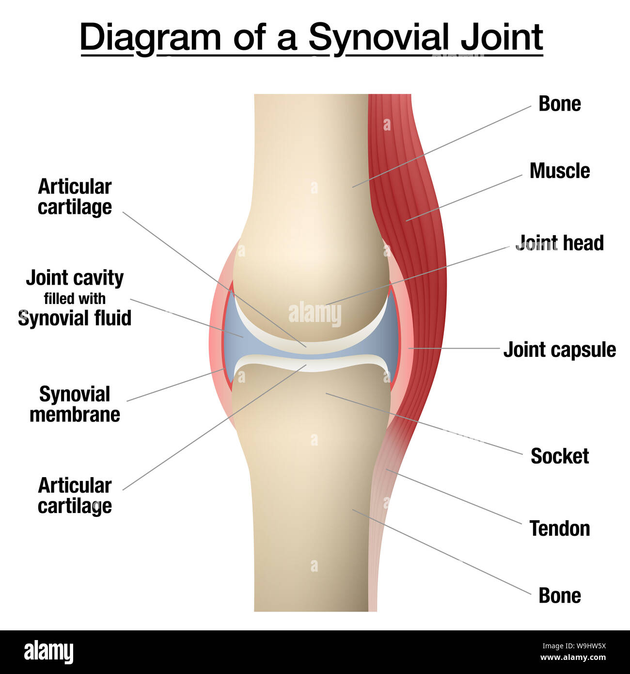
Synovial Joint Chart Labeled Anatomy Infographic With Two Bones Articular Cartilage Joint Cavity Synovial Fluid Muscle And Tendon Stock Photo Alamy from c8.alamy.com Tendonitis is the swelling of a tendon, which is a thick cord attaching a muscle to a bone. Human anatomy diagrams show internal organs. This will help you to understand the mechanism as well as the working. As these muscles contract and relax, they move skeletal bones to create movement of the body. Each of these muscles is a discrete organ constructed of skeletal muscle tissue, blood vessels, tendons, and nerves. Posted in diagrams , muscles | tagged human muscles , human muscles anatomy , muscle , muscle chart , muscle diagram , muscles , muscles anatomy. Almost every skeletal muscle works by pulling two or more bones either closer together or further apart. Tears of the achilles tendon can be tiny (microtears), or large, causing pain, swelling, and impaired movement.
For more anatomy content please follow us and visit our website:
They allow you to move and provide support for your upper body. Hand wrist u2013 graph diagram. This diagram depicts knee diagram tendons. The muscles that make up the quadriceps are the strongest and leanest of all muscles in the body. Download 1,037 muscle diagram male stock illustrations, vectors & clipart for free or amazingly low rates! Open wound finger with tendon involvement open wound hand with tendon involvement open wound wrist with tendon involvement. Just need a glimpse, leave your valuable advice let us know , and subscribe us! The knee joint, you need a perfectly labeled diagram of the knee. Knee tendon diagram / anatomy of the knee / ankle bones anatomy structure 10 photos of the ankle bones anatomy structure ankle tendons anatomy, elbow bones anatomy, hand bones anatomy, leg bones anatomy, shoulder bones anatomy, tibia anatomy, wrist bones anatomy, foot, ankle tendons anatomy, elbow bones anatomy, hand bones anatomy, leg bones anatomy. Leg tendons and ligaments diagram anatomy lower leg muscles tendons and ligaments leg tendons categories. Tendon, tissue that attaches a muscle to other body parts, usually bones. Muscle anatomy of the human arm, anterior view. Diagrams of the foot labeled.
Tendonitis is the swelling of a tendon, which is a thick cord attaching a muscle to a bone. The knee joint, you need a perfectly labeled diagram of the knee. Almost every skeletal muscle works by pulling two or more bones either closer together or further apart. Each circuit displays a distinctive voltage condition. The achilles tendon is the strongest and largest tendon in the body.
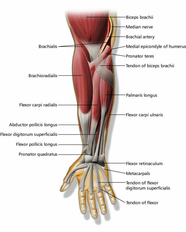
Muscles And Tendons Of Upper Extremity Labeled Eccles Health Sciences Library J Willard Marriott Digital Library from collections.lib.utah.edu Skeletal muscle diagram muscle fascia heart development types muscles fascia human body muscle and fascia heart cell fascia skeletal muscle cell anatomy muscular contraction. Attached to the bones of the skeletal system are about 700 named muscles that make up roughly half of a person's body weight. Joints act as pivot points for the movement of the bones. It should be noted that there are many more muscles in the body that are not addressed by this muscle anatomy diagram, however the muscles that are of primary interest from a fitness and exercise. Our latest youtube film is ready to run. Tendon sheaths, like tendons, are a type of muscles hand dorsal view labeled stock illustration 228843253 from image.shutterstock.com. New users enjoy 60% off. Tendons attach muscles to bones.
Knee tendon diagram / anatomy of the knee / ankle bones anatomy structure 10 photos of the ankle bones anatomy structure ankle tendons anatomy, elbow bones anatomy, hand bones anatomy, leg bones anatomy, shoulder bones anatomy, tibia anatomy, wrist bones anatomy, foot, ankle tendons anatomy, elbow bones anatomy, hand bones anatomy, leg bones anatomy.
See muscle contraction diagram stock video clips. Allows the action of raising the foot. We are pleased to provide you with the picture named hand nerve, tendon, and muscle anatomy.we hope this picture hand nerve, tendon, and muscle anatomy can help you study and research. Tendonitis is the swelling of a tendon, which is a thick cord attaching a muscle to a bone. It is the conjoined tendon of the gastrocnemius and the soleus muscles, and may have a small contribution from the plantaris. Tendons attach muscles to bones. Muscle anatomy atlas 12 photos of the muscle anatomy atlas , human muscles. Our latest youtube film is ready to run. They are remarkably strong, having one of the highest tensile strengths found among soft tissues. 19 photos of the knee tendon anatomy diagram and name chart. The majority of muscles in the leg are considered long muscles, in that they stretch great distances. This diagram depicts muscle in the body 744×1054 with parts and labels. If you tear the biceps tendon at the shoulder, you may lose some strength in your arm and have pain when you forcefully turn your arm from palm down to palm up.
They allow you to move and provide support for your upper body tendon diagram. See muscle contraction diagram stock video clips.

0 Comments:
Posting Komentar